
Digital health imaging
Redefining AI to revolutionise healthcare
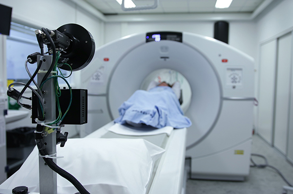
Under the theme of digital health imaging, we are developing next-generation AI technologies to revolutionise healthcare with the capacity to improve diagnostic efficiency and accuracy. With this, we aim to:
- bridge gaps between the increasing needs for medical practitioners and limited medical resources
- monitor disease progression at a subclinical level to inform personalised treatment planning and patient management
- further our understanding of disease and continue to pioneer innovations in healthcare and biotechnologies
- investigate more advanced AI solutions that support the shift from hospital-based to virtual-based care
We are a group of researchers across the University of Sydney, covering a broad range of multi-disciplinary research areas related to AI and healthcare. Under the umbrella of Digital Health Imaging, we are united by our close and productive multidisciplinary collaboration, a highly collaborative clinical research environment, and a strong link to industry partners.

Current projects

In this project, we will focus on cohesively developing advanced AI technologies to revolutionise imaging-based diagnosis of disease and extend our work towards commercial applications and clinical deployment. Specifically, we will first target three disease area cohorts where AI can greatly assist medical professionals: cancer (breast, brain, melanoma), lung diseases (e.g., COVID-19), and neurological conditions.

This MRFF project seeks to build a novel, hybrid AI learning ecosystem to generate clinically-relevant biomarkers of disease progression for the common, disabling neurological condition, multiple sclerosis (MS). The MSBASE-XNAT imaging repository and I-MED clinical radiology site data will respectively form the key components of a unique central-federated AI learning environment, yielding algorithms that, validated in a clinical neurology environment, will set a benchmark in diagnostic MS imaging; track subclinical progression of the disease; direct therapeutic strategy; and mine hitherto untapped quantitative imaging data.
This project involves close collaboration with Prof Michael Barnett and Dr Chenyu Tim Wang from the Faculty of Medicine and Health.

In this project, we will study the research area “explainable AI” that enables the outputs from the AI systems to exhibit a certain degree of explainable capability in order for clinical experts to better understand how AI systems diagnose diseases. The feedback from the clinical experts could in turn help us to develop better deep learning algorithms in the next round.

In this project, we will investigate how to introduce human intelligence in the loop when developing advanced AI methods, to progressively improve disease diagnosis results by taking advantage of both AI and human intelligence.

This project aims to exploit advanced AI technologies to enhance the quality of MRI data to make it comparable to that obtained from time consuming high-quality research protocols. The increased image data quality opens up the possibility of reliably applying advanced analysis methods to standard clinical data. The methods will be demonstrated in a number of neurological conditions.

Underpinning our leading AI research, we will expand on the AIS platform, designing and implementing a suite of digital tools to accelerate AI imaging research, better utilise the university’s hardware investments, securely collaborate with health and commercial partners, and provide a provenance trail for establishing trust.
Core research team
Professor Fernando Calamante is the Director of Sydney Imaging, the biomedical imaging Core Research Facility at the University of Sydney. He is Professor in the School of Biomedical Engineering, Faculty of Engineering. He is also Co-Director of the USyd/ANSTO joint node of the NCRIS-funded National Imaging Facility.
After he finished his BSc degree in Physics (with Honours) in Argentina, he went to study Magnetic Resonance Imaging (MRI) in the UK as a Chevening Scholar (The British Council), where he later carried out his PhD at University College London, and was subsequently appointed Lecturer in Biophysics (2000). He relocated to Australia in 2005, where he spent 12 years at The Florey Institute of Neuroscience and Mental Health and the University of Melbourne. Fernando joined the University of Sydney in 2018.
His main areas of research are the development of novel methods for Diffusion MRI, Perfusion MRI and brain connectivity, and their applications to neurology and neuroscience. He has gained international recognition for this work and his MRI methods have been adopted by many labs. For example, his MRtrix software for Diffusion MRI analysis is widely used worldwide and is one of the most popular tools in the field.
His research funding totals over $44M, including multiple ARC and NHMRC Project Grants and Fellowships, 2 NHMRC Program Grants, MRFF funding, and industrial funding.
Fernando has been elected to several leadership positions within the International Society for Magnetic Resonance in Medicine, including as its 2021-2022 President.
Dr Luping Zhou works on the interface of medical image analysis, machine learning, and computer vision. She obtained her PhD from ANU and got her post-doctoral training in UNC at Chapel Hill for computational medical image analysis. She is now a Senior Lecturer and ARC DECRA fellow in University of Sydney. A significant part of Luping’s research is image-based early diagnosis of mental disorders including the Alzheimer’s disease, ADHD, and epilepsy, etc. She developed a set of AI algorithms that advance the state-of-the-art work in brain image analysis. Luping’s current research is focused on medical image analysis with statistical graphical models and deep learning. Her research cares for significant problems of both scientific and commercial interests, such as dose-less medical imaging, medical image synthesis, segmentation and classification, and automated medical report generation, etc. Luping’s research has been supported by ARC DP, ARC DECRA, and Microsoft AI for Accessibility grant, etc.
Dr Ryan Sullivan is a Product Specialist in Characterisation in the Research Technology Group and an Honorary Associate in BME. He has a BS (Biophysics) from Wake Forest University and a PhD in medical implant fabrication and characterisation from UoW He leads the Australian Imaging Service (AIS), a national platform of 11 Australian universities and medical research institutes in partnership with the National Imaging Facility and the ARDC. He co-developed the NCRIS 5-year Australian Microscopy eResearch Roadmap. As a CI he has attracted $7.6M in project funding in the past 2 years ($1.85M as Lead/Sole CI), with an additional $1.1M co-investment from partners; and his leadership in big data and eResearch has been recognised by 8 invited national lectures in 2019.
Our partners
We work with eHealth NSW on secure data for clinical research, and through our Australian Imaging Service, work with eHealth Queensland, Victoria Health, and WA Health.
We also work with private sites such as I-MED Radiology Network and Melanoma Institute Australia with our clinical colleagues.